What Is Ptosis (Droopy Eyelid)?
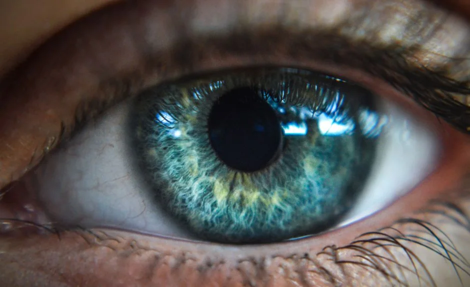
Ptosis — pronounced “TOE-sis,” from the Greek for “falling” — is the abnormal drooping of one or both upper eyelids below their normal resting position.
Clinically, it is defined as a marginal reflex distance (MRD1, the gap between the corneal light reflex and the upper eyelid margin) of less than the normal 4–5 mm. When the lid descends over the pupil, it reduces the upper visual field; the brain compensates by recruiting the frontalis (forehead) muscle to hold the brow elevated, a habit that underlies many cases of tension headache and eye strain attributed to ptosis.
The condition ranges from mild — a barely perceptible asymmetry — to severe, where the lid fully or nearly fully covers the pupil. Both functional impairment and the aesthetic impact of a persistently tired expression are legitimate reasons to seek evaluation.
Causes and Types of Ptosis
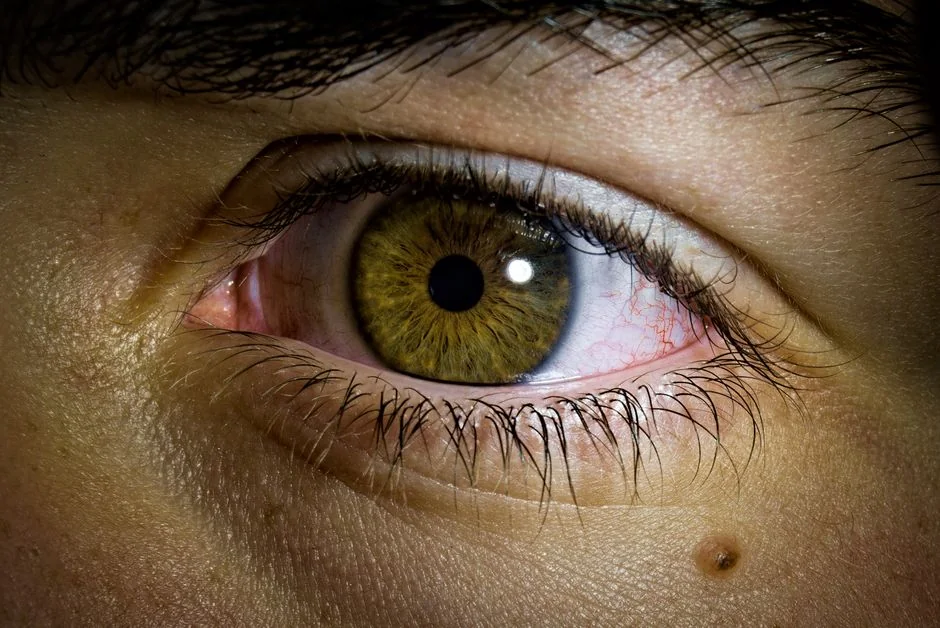
Aponeurotic (Involutional) Ptosis — the Most Common Form
In adults, the overwhelming majority of new-onset ptosis is aponeurotic, also called involutional or senile ptosis.
The levator aponeurosis — the tendon connecting the upper eyelid’s main elevator muscle to the tarsal plate — gradually detaches, stretches, or thins with age. The muscle itself remains functional; it is the mechanical connection to the eyelid that has failed.
Common triggers beyond natural ageing include chronic contact lens wear (repeated upward traction on the lid during insertion and removal), habitual eye rubbing, and previous ocular procedures such as cataract surgery, after which one published series documented a ptosis incidence of 7.3%.
Other Types at a Glance
- Congenital ptosis — present from birth, caused by incomplete levator muscle development; surgical correction in childhood prevents amblyopia.
- Myogenic ptosis — muscle-disease related (myasthenia gravis, oculopharyngeal muscular dystrophy); must be diagnosed and stabilised before surgical planning.
- Neurogenic ptosis — caused by nerve dysfunction, including third cranial nerve palsy or Horner syndrome; systemic investigation is required first.
- Mechanical ptosis — the eyelid is weighed down or tethered by excess skin, a tumour, or scarring.
Identifying the underlying cause is essential: technique selection and expected outcome differ substantially across types.
How Ptosis Correction Surgery Works
The surgical goal is to restore the upper eyelid margin to an appropriate height — generally 1–2 mm below the upper corneal limbus — while achieving symmetry with the opposite eye. The technique is chosen based primarily on the degree of ptosis and the residual excursion (movement) of the levator muscle.
Levator Aponeurosis Advancement (External Approach)
For aponeurotic ptosis with preserved levator function of 8 mm or more, levator advancement is the standard procedure. A small incision is made in the natural eyelid crease; the surgeon locates the disengaged or attenuated aponeurosis and reattaches it to the tarsal plate. Because the underlying muscle is intact, this restores normal anatomy. Clinical studies suggest strong outcomes: one prospective study of 810 eyelids in 450 patients reported a success rate of 94.9%, with mean MRD1 improving from 1.7 mm pre-operatively to 3.7 mm at six-month follow-up.
Müller Muscle-Conjunctival Resection (Internal Approach)
For mild ptosis with good levator function (≥ 10 mm), a posterior (internal) approach avoids an external skin incision entirely. The surgeon accesses Müller’s muscle — a thin involuntary smooth muscle beneath the levator — through the inner eyelid surface. Candidates are typically identified by a positive phenylephrine test: if a topical sympathomimetic drop temporarily raises the lid by 2 mm or more in clinic, the Müller muscle is likely responsive to surgical resection.
Frontalis Suspension
When levator function is severely compromised — typically less than 4–5 mm of excursion — neither the levator nor Müller’s muscle can be relied upon to elevate the lid reliably. In these cases, the eyelid is suspended from the frontalis muscle using a sling of silicone rod, autologous fascia lata, or synthetic material, so that voluntary brow elevation opens the eye.
| Technique | Best Indication | Levator Function | Incision Approach |
|---|---|---|---|
| Levator aponeurosis advancement | Aponeurotic / involutional ptosis | Good (≥ 8 mm) | External (eyelid crease) |
| Müller muscle-conjunctival resection | Mild ptosis, phenylephrine-positive | Good (≥ 10 mm) | Internal (conjunctival) |
| Frontalis suspension | Severe ptosis, poor levator function | Poor (< 5 mm) | Small stab incisions (brow and lid) |
Expected Results and Recovery
What the Evidence Shows
For aponeurotic ptosis — the type most commonly seen in adults seeking surgery — clinical reports indicate that levator-based repair achieves high initial success rates.
The longer-term picture is that some degree of recurrence is possible: a 2025 multicentre study following 152 eyes over a median of approximately 3.2 years found an overall recurrence rate of 23%, with aponeurotic ptosis recurring in 19.5% of cases versus 26.7% for non-aponeurotic forms.
Crucially, the study concluded that ptosis aetiology — rather than any preoperative measurement or choice of surgical technique — was the primary predictor of long-term durability. Patients with aponeurotic ptosis can therefore expect the most favourable and predictable outcomes. Undercorrection — where the eyelid remains slightly lower than the target — occurs in an estimated 10–15% of cases and can be addressed with a minor revision.
Recovery Timeline
- Days 1–3: Swelling and bruising are expected; cold compresses and head elevation help. Most patients rest at home.
- Days 7–10: External sutures removed. Swelling begins to resolve noticeably.
- 2–4 weeks: Presentable for most work and daily activity; makeup may usually be resumed.
- 3–6 months: Final result as residual swelling fully settles and the eyelid finds its resting height.
Ptosis Surgery Cost in Tokyo: Insurance vs. Self-Pay
When Japan’s National Health Insurance Applies
Ptosis correction in Japan qualifies for national health insurance (健康保険) coverage when a physician documents functional visual impairment — most commonly when the MRD1 is at or below 2 mm, or when the upper visual field is measurably restricted. The surgery must be performed to restore function, not for aesthetic improvement alone.
When the insurance criteria are met, the surgical fee for levator advancement is governed by the national fee schedule (診療報酬点数表). With a standard 30% patient co-payment, the surgery itself costs approximately ¥22,000 per eye; consultation fees and post-operative medications are billed separately. Patients holding a valid Japanese health insurance card (保険証) — including enrolled foreign residents — are eligible.
Cosmetic (Self-Pay) Ptosis Correction
When surgery is sought for aesthetic reasons without a qualifying functional impairment, it is classified as self-pay (自由診療). Costs vary by clinic, technique, and whether bilateral correction is required. Please see the clinic’s official price page or contact us for a current quote. For an accurate assessment of whether your case may qualify for insurance, please book a consultation.
Have a question about this treatment?
Message us on LINE or WhatsApp — our English-speaking team usually replies the same day.
What to Expect: Before, During, and After Surgery
Pre-operative Assessment
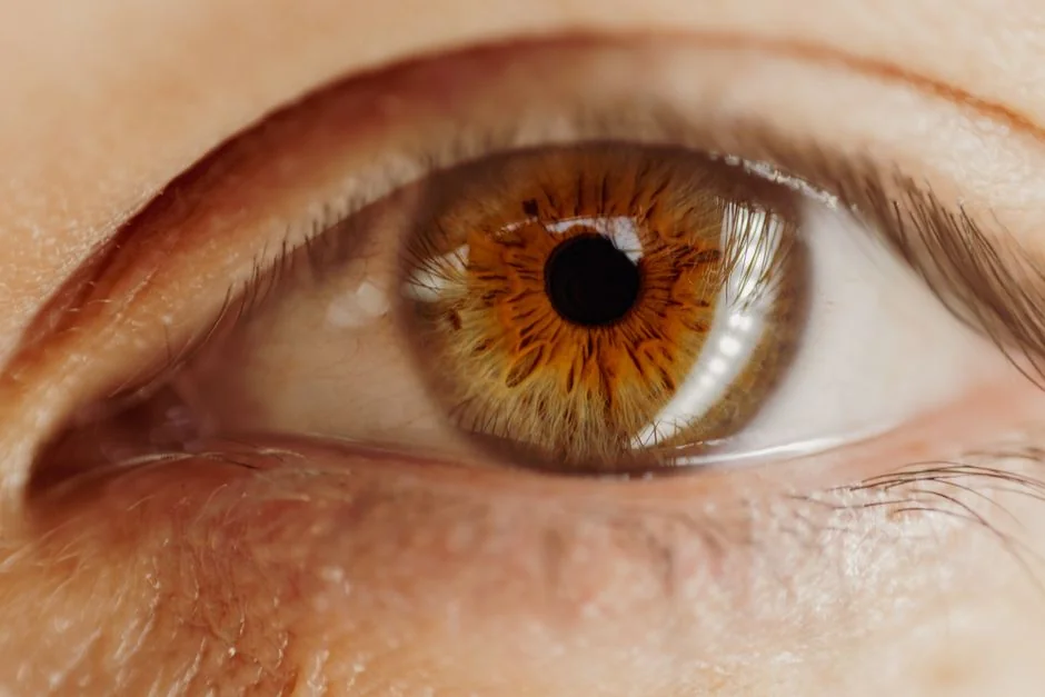
A thorough consultation includes measurement of MRD1 and levator excursion, assessment of eyelid crease height, corneal sensation testing, and evaluation for dry eye. Photographs are taken in primary gaze and looking up. Blood-thinning medications — including aspirin, NSAIDs, and certain supplements — are usually paused for one to two weeks prior to surgery on the surgeon’s instruction. Patients should disclose all medications, including topical eye drops.
During Surgery
Ptosis correction is performed under local anaesthesia, typically with optional mild intravenous sedation, as a day-case procedure lasting approximately 30–90 minutes. For external levator advancement, the surgeon will often ask the patient to open their eyes briefly mid-procedure while awake — this intraoperative check allows real-time adjustment of eyelid height and symmetry, which is difficult to assess under general anaesthesia.
Post-operative Care
Antibiotic eye drops or ointment are prescribed for the first week. Patients are advised to avoid strenuous exercise, swimming, and heavy lifting for approximately three weeks. Sleeping with the head slightly elevated reduces post-operative oedema. Sun exposure should be minimised during the healing phase. Follow-up appointments at one week and one month are standard.
Who Is a Good Candidate?
Patients most likely to benefit include those who:
- Have a measurable reduction in eyelid height (MRD1 below the normal range of 4–5 mm) causing functional limitation or significant aesthetic concern
- Have good overall ocular health and adequate tear film (dry eye is assessed and managed before proceeding)
- Hold realistic expectations about the gradual resolution of swelling over several months and the possibility of minor asymmetry
Surgery is approached with additional caution in patients who have:
- An active autoimmune or neuromuscular condition such as myasthenia gravis (must be excluded before surgery)
- Severe dry eye disease, which incomplete eyelid closure after surgery can exacerbate
- Thyroid-associated orbitopathy in an active or recently active phase
- Significant scarring from prior eyelid procedures that may complicate dissection
Common Misconceptions About Ptosis Correction
“Ptosis surgery and double eyelid surgery are the same thing”
This confusion is understandable, because both involve the upper eyelid and are sometimes performed together. They are, however, distinct procedures addressing different anatomy. Double eyelid surgery (重瞼術) creates or refines the upper eyelid fold by forming an adhesion between the skin and the underlying tarsal plate or levator — it does not change the height of the eyelid margin. Ptosis correction physically elevates the eyelid margin by reattaching or tightening the levator mechanism. Performing a double eyelid procedure when ptosis is the true underlying problem is a recognised cause of unsatisfactory outcomes that may later require revision.
“Ptosis is only a problem for older adults”
While involutional aponeurotic ptosis becomes more prevalent with age, the condition affects all age groups. Chronic contact lens wear — particularly with rigid lenses worn for many years — is a well-documented cause in younger adults, as repeated mechanical traction on the upper lid stretches the aponeurosis by the same mechanism as ageing. Congenital ptosis is, by definition, present from birth. Any patient presenting with a persistently low upper eyelid margin warrants an assessment regardless of age.
Where to Get Ptosis Correction in Tokyo
Within our clinic group, both BIOTOPE CLINIC (Shirokanedai, Minato-ku) and Kojimachi Dermatology & Plastic Surgery Clinic (Ichigaya, Chiyoda-ku) offer English-language consultation with Dr. Jun Karibe’s team. Cosmetic ptosis correction is handled at BIOTOPE CLINIC in Shirokanedai; insurance-covered functional ptosis cases are assessed and operated at Kojimachi Dermatology & Plastic Surgery Clinic in Ichigaya. Bookings and enquiries in English: LINE or WhatsApp.
At BIOTOPE CLINIC in Shirokanedai, Dr. Karibe’s team frequently sees patients whose concern spans both the functional heaviness of the upper eyelid and its aesthetic effect. In such cases, a careful clinical assessment determines whether insurance criteria are met; if not, a personalised cosmetic treatment plan is discussed. Patients wishing to combine ptosis correction with related eyelid procedures — such as double eyelid surgery using the suture method, which is available within the clinic group — are welcome to raise this during consultation.
Frequently Asked Questions
- Q. Is ptosis correction painful?
- The procedure is performed under local anaesthesia, so the operative site is fully numbed before any incision. Most patients experience a brief sting from the anaesthetic injection and no pain during surgery itself. Post-operative discomfort is generally mild and managed with standard over-the-counter analgesics for a few days.
- Q. How long do the results last?
- For aponeurotic ptosis, results are typically long-lasting. Clinical data suggest that over a median follow-up of approximately three years, recurrence occurs in roughly 19–20% of aponeurotic cases. If ptosis does return, a revision procedure using a similar technique is usually feasible, and long-term outcomes remain good for this category of patient.
- Q. Can I have ptosis correction if I have had eyelid surgery before?
- In many cases, yes — though previous surgery creates scar tissue that can complicate dissection and may require a modified approach. The surgeon will assess the degree of scarring and the integrity of the remaining levator mechanism at consultation. Results following revision ptosis surgery are generally good, but detailed pre-operative planning is essential.
- Q. Will my eyes look symmetrical after the operation?
- Surgeons aim to achieve eyelid heights within 0.5–1 mm of each other; asking the patient to open their eyes briefly mid-procedure while under local anaesthesia allows real-time fine-tuning. A small degree of asymmetry is considered a normal outcome in eyelid surgery and is rarely functionally significant. Perfect symmetry cannot be guaranteed.
- Q. Does Japan’s national health insurance cover ptosis surgery for foreign nationals?
- Japan’s national health insurance schemes (健康保険 and 国民健康保険) cover foreign nationals who are enrolled — this typically applies to residents on long-term visas who have registered with their municipal office. Short-term visitors holding a tourist or business visa are not enrolled and pay self-pay rates. If you are enrolled, please bring your insurance card (保険証) to the consultation.
Have a question about this treatment?
Message us on LINE or WhatsApp — our English-speaking team usually replies the same day.
Summary
Ptosis correction surgery restores the upper eyelid to an appropriate height by repairing or compensating for the levator mechanism that lifts it. The most common form in adults — aponeurotic ptosis — is well-suited to levator advancement, with clinical studies reporting success rates of approximately 94–95% at six months. Longer-term data indicate that some recurrence is possible over years, particularly in non-aponeurotic types; aponeurotic ptosis carries the most favourable prognosis. In Japan, the procedure can be covered under national health insurance when functional visual impairment is documented, making it more accessible than comparable surgery in many other countries. Where no qualifying impairment exists, cosmetic self-pay correction is available.
If you have questions about ptosis correction, both BIOTOPE CLINIC (Shirokanedai, Minato-ku) and Kojimachi Dermatology & Plastic Surgery Clinic (Ichigaya, Chiyoda-ku) offer English-language consultation with Dr. Karibe and his team. Cosmetic ptosis correction is primarily handled at BIOTOPE CLINIC in Shirokanedai; insurance-covered functional ptosis cases are assessed at Kojimachi Dermatology & Plastic Surgery Clinic in Ichigaya. English enquiries: LINE or WhatsApp.
24/7 Consultation & Booking
Book a Consultation or Treatment
Our English-speaking team responds via LINE or WhatsApp — usually the same day.
References
- Mild to moderate blepharoptosis correction: outcomes of levator aponeurosis posterior layer plication. PLOS ONE. 2020.
- Long term risk of recurrence of ptosis repair: implications for surgical counseling and follow-up. Frontiers in Ophthalmology. 2025.
- American Academy of Ophthalmology EyeWiki. Aponeurotic Blepharoptosis (Ptosis). Accessed November 2026.
- A review of acquired blepharoptosis: prevalence, diagnosis, and current treatment options. Eye (London). 2021.
Related Articles
You may also find these articles useful.
- Double Eyelid Surgery Tokyo: English-Speaking Clinic Guide
- Epicanthoplasty in Tokyo: Inner Eye Corner Surgery
- Under Eye Bags Surgery Tokyo: Transconjunctival Guide
- Under-Eye Dark Circles Tokyo: Filler Injections vs Surgery
- Finding an English-Speaking Plastic Surgeon in Tokyo
SUPERVISED BY

Dr. Jun Karibe
MD
Director
Education & Career
Certifications
Awards
MEDICAL REVIEW