What Is Filler Vascular Safety Screening?
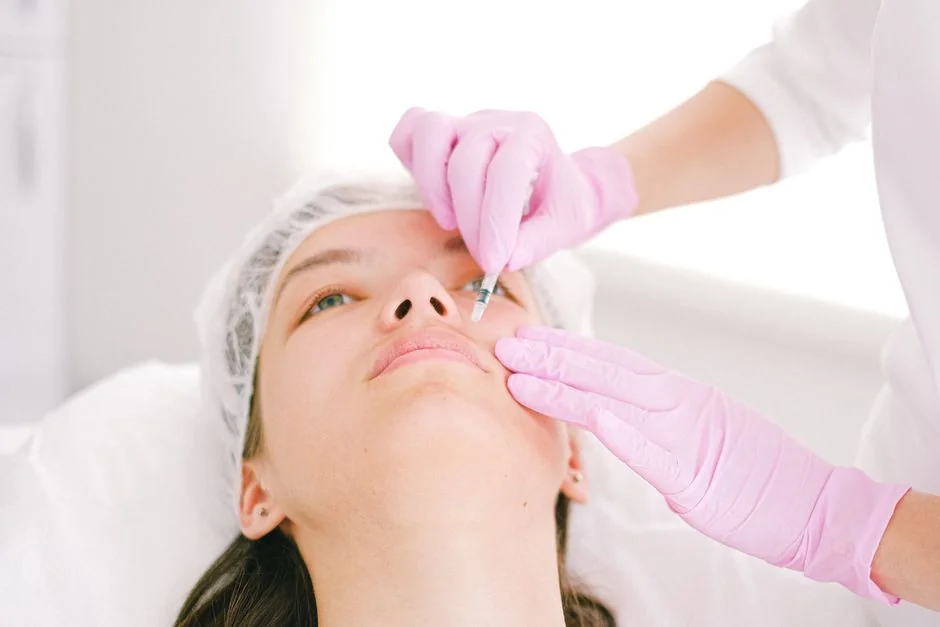
Vascular safety screening for filler is the clinical step of identifying blood vessel positions, depths, and flow patterns in a treatment area before the injection begins, so that the needle or cannula path can avoid them; the most accurate tool for this is high-resolution Doppler ultrasound, which visualises vessels in real time without radiation.
Dermal fillers — most commonly hyaluronic acid (HA) — are injected into specific tissue planes to restore volume or smooth contour. When an injection inadvertently enters a blood vessel, the filler particle acts as an embolus, obstructing downstream blood flow. The clinical outcome depends on which vessel is affected and how quickly treatment is initiated: the range spans from bruising and swelling at the mild end, through full-thickness skin necrosis, to permanent vision loss in the most severe cases.
A 2024 cumulative review published in Aesthetic Surgery Journal documented 511 cases of filler-related blindness spanning 1906 to March 2023, with 365 of those cases published in just the five years between 2018 and 2023 alone
— a sharp increase compared with all prior decades combined. Hyaluronic acid was the filler material in 79.6% of recent cases, and only 6.0% of affected patients experienced complete vision recovery.
These figures make clear that pre-treatment vascular assessment is not a precautionary luxury; it is a clinically meaningful intervention, particularly in anatomically complex zones.
Vessel positions vary meaningfully between individuals. A supratrochlear artery that runs centrally in one patient may run laterally in another; a dorsal nasal artery at 3 mm depth in one case may sit at 1.5 mm in another. This individual variability makes textbook anatomical knowledge an imprecise guide for every patient, every session.
How Ultrasound Mapping Works — and Which Zones Require It Most

Pre-treatment ultrasound mapping uses a linear high-frequency probe — a minimum of 15 MHz is recommended by the 2025 international expert consensus — combined with colour Doppler to display each vessel’s position, depth, and flow direction before the injection begins, producing an individual vascular map the practitioner uses to plan depth, angle, and entry point.
Three imaging modes work in combination. B-mode (greyscale) reveals soft tissue architecture and identifies prior filler deposits, which alter local anatomy and are invisible on the surface.
Colour Doppler overlays blood-flow direction onto the greyscale image: arteries and veins appear as coloured signals that immediately confirm their precise locations. Spectral Doppler then analyses flow velocity and resistance, distinguishing high-pressure arteries from veins — an important distinction because arterial occlusion carries far greater complication risk than venous involvement.
A 2025 modified Delphi consensus published in the Journal of Plastic, Reconstructive & Aesthetic Surgery, drawing on fifteen experts across seven specialties and eleven countries, concluded that ultrasound imaging prior to injection is mandatory in areas carrying a high risk of vascular adverse events. The expert group also recommended that any existing filler deposits be identified by ultrasound before additional product is introduced — a step not possible through surface inspection alone.
High-Risk Zones: Where Filler Accidents Concentrate
Clinical data consistently identify five facial zones as carrying the highest risk of serious vascular events. A 2025 systematic review in Cureus, analysing fourteen studies from 2009 to 2022, found that the glabella, nose, and nasolabial folds were most commonly implicated in occlusion cases. A parallel literature review of vision-loss cases traced the injection sites to the nose (36.5%), glabella (27.0%), and forehead (20.2%) most frequently.
| Facial Zone | Key Vessels at Risk | Potential Complication | Ultrasound Priority |
|---|---|---|---|
| Nose | Dorsal nasal artery, lateral nasal artery | Skin necrosis, vision loss | High |
| Glabella | Supratrochlear artery, supraorbital artery | Skin necrosis, vision loss | High |
| Forehead | Supraorbital artery, superficial temporal branches | Vascular occlusion, vision loss | High |
| Temple | Superficial temporal artery | Occlusion, arteriovenous fistula | High |
| Nasolabial fold | Angular artery, facial artery | Vascular occlusion, skin necrosis | Medium–High |
| Lips | Superior and inferior labial arteries | Vascular occlusion, bruising | Medium |
The anatomical reason that the nose and glabella carry disproportionate severity is the arterial network they share with ocular circulation. The dorsal nasal and angular arteries connect through the ophthalmic artery to the central retinal artery. A bolus of filler entering the wrong plane in these zones can travel retrogradely along this route in seconds — faster than emergency intervention can reach.
What to Expect at Your Appointment, and What It Costs in Tokyo
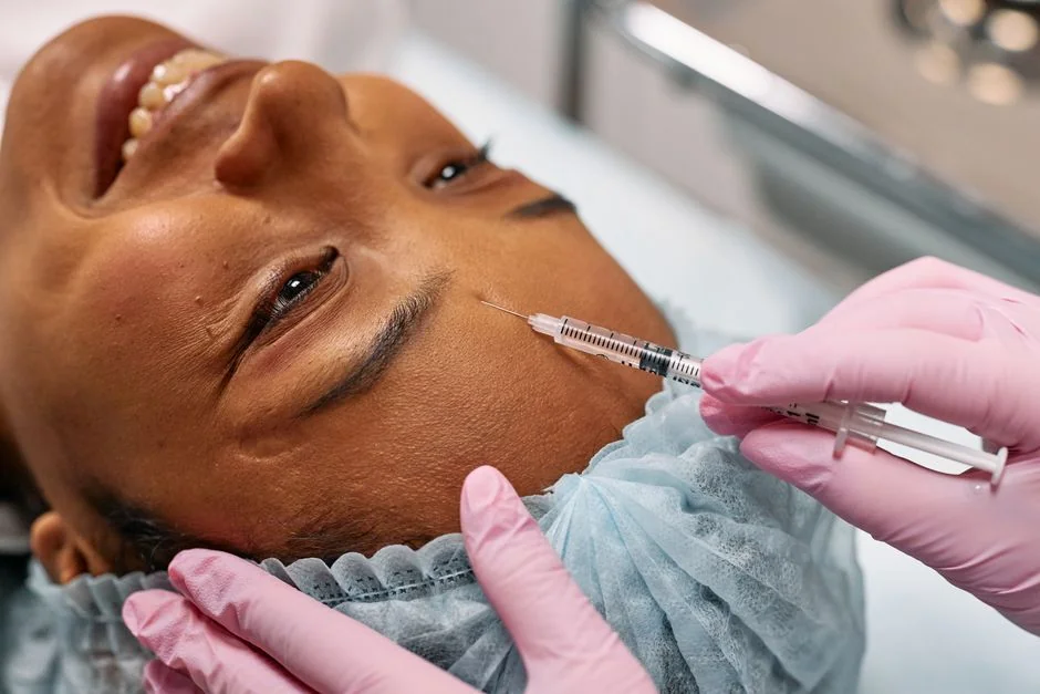
A filler appointment that includes pre-treatment ultrasound mapping adds a brief vascular assessment stage before injection — typically five to ten minutes per treatment zone — after which the practitioner proceeds with an individualised vessel map rather than a generic anatomical template; total cost in Tokyo reflects the filler product, clinical time, and whether real-time guidance versus mapping alone is used.
Before the Injection
At consultation, the practitioner reviews your medical history — including prior filler treatments, blood-thinning medications, and any known reactions — and discusses the planned treatment sites. For high-risk zones, a vascular mapping session is scheduled, either immediately before injection or as a preliminary appointment. Patients who have had prior filler in the same area benefit particularly from pre-treatment imaging: existing HA deposits displace local vessels in ways that cannot be predicted from the surface.
During the Mapping Session
Ultrasound gel is applied to the treatment area. The probe moves across the skin as the practitioner observes vessel positions, depths, and trajectories in real time. Any vessels within the planned injection path are noted and factored into the injection plan. For the highest-risk sites — particularly liquid rhinoplasty and glabellar correction — real-time ultrasound guidance during the injection itself may be recommended in addition to pre-treatment mapping.
After Treatment: Warning Signs to Know
All patients should know the early warning signs of vascular compromise: blanching of the skin immediately after injection, disproportionate pain beyond expected tenderness, or any sudden change in vision. These require immediate assessment, not observation at home. Routine bruising and swelling are normal and typically resolve within days. Before leaving the clinic, confirm that hyaluronidase is available on-site and that the team has a documented occlusion response protocol.
Cost in Tokyo
Hyaluronic acid filler pricing in Tokyo varies by product brand, volume, and treatment complexity. Pre-treatment ultrasound assessment may be included within the consultation fee at some clinics, or charged separately at others. Patients planning treatment in high-risk zones — the nose, glabella, forehead, or temples — should approach vascular assessment as part of the clinical baseline, not an optional add-on. Please see the clinic’s official price page or contact us for a current quote. For an accurate quote, please book a consultation.
Have a question about this treatment?
Message us on LINE or WhatsApp — our English-speaking team usually replies the same day.
Who Is a Good Candidate — and Who Should Reconsider?

Most healthy adults seeking facial volume restoration with hyaluronic acid filler are suitable candidates; those planning treatment in high-risk anatomical zones, those with prior filler deposits in the same area, or those on anticoagulant medication benefit most from a formal ultrasound vascular assessment before treatment proceeds.
Suitable candidates include adults in good general health addressing volume loss in the cheeks, temples, nasolabial folds, lips, or under-eye area. Patients seeking nose filler (liquid rhinoplasty) or glabellar correction are advised to have a full vascular mapping session before the injection, given the direct anatomical links to ophthalmic circulation described above. Hyaluronic acid filler treatment is available at BIOTOPE CLINIC in Shirokanedai — the team is happy to explain the specific screening protocol for your area of interest during an English-language consultation with Dr. Karibe.
Relative contraindications include active skin infection or inflammation at the injection site, blood-thinning medication that cannot be safely paused, autoimmune conditions affecting wound healing, and unrealistic expectations about the degree of change achievable in a single session. Pregnancy and breastfeeding are contraindications for elective cosmetic procedures.
What We See Most Often at BIOTOPE CLINIC in Shirokanedai
At BIOTOPE CLINIC in Shirokanedai, a portion of patients seeking filler consultations arrive having received prior treatments at other providers — sometimes across multiple sessions over several years. In these cases, ultrasound assessment before retreatment is especially relevant, because pre-existing HA deposits can compress or displace local vessels in ways that are not apparent from the surface. It is also common for patients to be uncertain about which filler material or volume was used in prior treatments, making imaging-based identification of residual product a useful clinical baseline before introducing additional volume into a high-risk zone. This is an operational reality at any specialist aesthetic clinic that sees patients with complex filler histories.
Common Misconceptions About Filler Vascular Safety
Misconception: “Using a cannula instead of a needle eliminates the risk.”
Cannulas meaningfully reduce but do not eliminate vascular occlusion risk. A JAMA Dermatology study reviewing 1.7 million syringes across 370 board-certified dermatologists found that cannula injections carried 77.1% lower odds of occlusion compared with needles — a clinically significant reduction, but not a zero-risk outcome. The same dataset showed that practitioners with more than five years’ experience demonstrated 70.7% lower odds of occlusion than those with less experience, confirming that instrument choice alone does not account for the full safety profile. Pre-treatment vascular mapping is advisable in high-risk zones regardless of which instrument is used.
Misconception: “I have had filler with no complications before, so my anatomy is safe.”
Prior uneventful treatment means the injection did not enter a vessel on that occasion — it does not mean the anatomy is permanently navigable at the same coordinates. Prior filler deposits alter local tissue architecture, age-related volume changes shift tissue planes over time, and an absence of complications in lower-risk zones does not predict safety in higher-risk ones. Each high-risk zone benefits from assessment on its own merits at each treatment episode.
Frequently Asked Questions
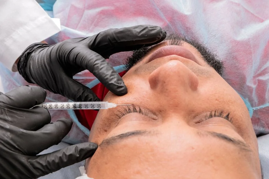
The questions patients most commonly raise before filler consultations in Tokyo cover the availability of ultrasound mapping, what happens if an occlusion occurs despite precautions, appointment logistics, and English-language access — each is addressed below with specific clinical detail.
- Q. Is pre-treatment ultrasound mapping available at most filler clinics in Tokyo?
- Adoption varies. The 2025 international Delphi consensus recommends it as mandatory for high-risk zones, but not all clinics have incorporated this into standard practice. Patients should ask explicitly whether vascular mapping is part of the protocol for their planned treatment site, and what probe frequency the clinic uses. BIOTOPE CLINIC in Shirokanedai incorporates vascular assessment as part of the consultation process for high-risk sites.
- Q. What happens if a vascular occlusion occurs despite pre-treatment screening?
- Speed of response is the critical variable. For hyaluronic acid fillers, hyaluronidase — an enzyme that dissolves HA — is injected to restore blood flow; the 2025 Cureus systematic review found it achieved partial or complete recovery in 84.2% of HA-related occlusions. Any practitioner offering filler should have hyaluronidase on-site and a clear occlusion protocol, and patients should be informed of warning signs before leaving the clinic.
- Q. If I have had filler with no complications before, do I still need screening at my next appointment?
- Prior uneventful treatment is not a substitute for a current vascular assessment, particularly if the new treatment targets a different or higher-risk zone, or if prior filler has been deposited in the same area. Vessel position and tissue architecture change over time, and existing filler deposits can alter local anatomy in ways that affect injection planning for subsequent sessions.
- Q. I am an expat in Tokyo — can I book a consultation in English?
- Yes. Both BIOTOPE CLINIC in Shirokanedai and Kojimachi Dermatology & Plastic Surgery Clinic in Ichigaya offer English-language consultations. Filler treatment and vascular safety screening are handled at BIOTOPE CLINIC in Shirokanedai. English enquiries are available via LINE or WhatsApp.
Have a question about this treatment?
Message us on LINE or WhatsApp — our English-speaking team usually replies the same day.
Where to Get Filler Vascular Safety Screening in Tokyo
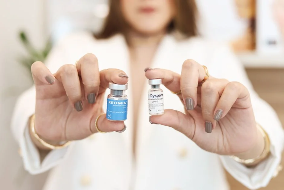
Within Dr. Jun Karibe’s clinic group, both BIOTOPE CLINIC (Shirokanedai, Minato-ku) and Kojimachi Dermatology & Plastic Surgery Clinic (Ichigaya, Chiyoda-ku) offer English-language consultation with Dr. Karibe’s team; filler injections and pre-treatment vascular safety screening are handled at BIOTOPE CLINIC in Shirokanedai, where Dr. Karibe personally consults.
Kojimachi Dermatology & Plastic Surgery Clinic in Ichigaya is the group’s main office, focused on insurance-covered dermatology — eczema, acne, rash management, general skin conditions — and general aesthetic plastic surgery. Patients who are uncertain which clinic to attend are welcome to reach out in English through either branch, and the team will advise on the most appropriate referral.
Vascular complications from hyaluronic acid filler injection are preventable with the right pre-treatment assessment. High-resolution Doppler ultrasound mapping, now supported by an international expert consensus as mandatory for high-risk zones, allows practitioners to tailor the injection plan to each patient’s vascular anatomy. The nose, glabella, forehead, and temple are the sites warranting the greatest care; cannula use reduces risk in these areas but does not eliminate it; and the speed with which a potential occlusion is recognised and treated determines the final clinical outcome.
If you have questions about filler vascular safety screening in Tokyo, both BIOTOPE CLINIC (Shirokanedai, Minato-ku) and Kojimachi Dermatology & Plastic Surgery Clinic (Ichigaya, Chiyoda-ku) offer English-language consultation with Dr. Karibe and his team. Filler treatment and vascular safety screening are handled at BIOTOPE CLINIC in Shirokanedai. English enquiries: LINE or WhatsApp.
24/7 Consultation & Booking
Book a Consultation or Treatment
Our English-speaking team responds via LINE or WhatsApp — usually the same day.
References
- Alam M, Kakar R, Dover JS, et al. Rates of Vascular Occlusion Associated With Using Needles vs Cannulas for Filler Injection. JAMA Dermatology. 2021;157(2):174–180.
- Chakhachiro A, Waseem M. Risk Factor Analysis for Vascular Occlusions After Dermal Filler Injections: A Systematic Review and Meta-Analysis. Cureus. 2025.
- Doyon VC, Liu C, Fitzgerald R, et al. Update on Blindness From Filler: Review of Prognostic Factors, Management Approaches, and a Century of Published Cases. Aesthetic Surgery Journal. 2024;44(10):1091–1102.
- Kato JM, Matayoshi S. Visual loss after aesthetic facial filler injection: a literature review on an ophthalmologic issue. Arquivos Brasileiros de Oftalmologia. 2022.
- Velthuis PJ, Alfageme F, Cartier H, et al. The use of ultrasound imaging in aesthetic injectables: A modified Delphi consensus. Journal of Plastic, Reconstructive & Aesthetic Surgery. 2025;104:33–37.
Related Articles
You may also find these articles useful.
- Hyaluronidase in Tokyo: A Guide to Dissolving HA Filler
- When to Dissolve HA Filler: Signs You Need Hyaluronidase
- Under Eye Filler Tokyo: Risks, Benefits & Costs
- Dermal Filler Tokyo: Price Guide & Clinic Selection Tips
- Lip Filler Tokyo: Cost, Treatment Guide & Results
SUPERVISED BY

Dr. Jun Karibe
MD
Director
Education & Career
Certifications
Awards
MEDICAL REVIEW